Friday, September 20, 2013

Have More Kids And Breastfeed Longer To Reduce Breast Cancer Risks

By Shahfizal Musa
Pix Ismail Ibrahim
KUALA LUMPUR, 21 Jun 201 - Women who have more children and breastfeed them for a long period of time can reduce the risk of getting breast cancer.
A pathologist at The National University of Malaysia (UKM), Prof Dr Aishah Md Ali said breast cancer can be reduced by half in developed countries by just having more children and breastfeeding their babies for some time.
Giving her inaugural lecture on ‘Women, Tumor and Cancer, the Pathological Perspective’  she explained in great detail about cancers that affect women.
Prof Aishah has written a book on the subject and had also presented more than 130 papers in various conferences around the world.
Though Malaysia is moving towards the developed nation status it can still significantly reduce the number of such cases if Malaysian women are to practice the risk mitigation methods.
She said those methods had been prescribed in Islam which were later discovered by western researchers.
In Malaysia cancer is the third highest cause of death in hospitals with breast cancer taking the lead, followed by cervix, colon, ovarian, leukemia and lung cancer.
Risks faced by the various racial groups in Malaysia have to be assessed separately as each race have their own lifestyles and their risk has to be assessed separately in order to give the best treatment possible.
She quoted a local study that evaluated the risks of cancer in the three major races, Malays, Chinese and Indians.  
 
The study shows that the risk of breast cancer is highest among Chinese and lowest for the Malays that is 1 in 16 chance for the Chinese to get breast cancer, 1 in 17 for the Indians and 1 in 28 chance for the Malays.
Ethnic
Breast Cancer Risk
Chinese
        1:16
Indian
        1:17
Malay
        1:28
The Chinese also faces a higher risk than the rest when it comes to cervical cancer. The life expectancy is reduced from one to 40 Chinese suffering from cervical cancer. For the Indians the risk is 1 in 50 of a reduced life expectancy, while for the Malays the risk of life expectancy reduction is only 1 in 111.
Ethnic
Life Expectancy Risk
Chinese
        1:40
Indian
        1:50
Malay
        1:111
For cervical cancer 80 percent of the dire cases are found in developing countries as severe cases in developed countries have been reduced due to the use of screening methods enabling early detections.
Present at the lecture was the Deputy Vice Chancellor (Academic and International Affairs), Professor Dato' Ir Dr Othman A. Karim; Dean of the Faculty of Medicine, Professor Dato’ Dr Raymond Azman Ali, academics, physicians and students.

UKMMC First Hospital in Asia to have safer thyroid surgery

By Shahfizal Musa
Pix Izwan Azman
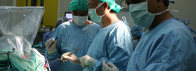
KUALA LUMPUR 22 July 2013 – The National University of Malaysia Medical Centre is the first Hospital in Asia to use the latest technique in thyroid surgery which drastically reduces risk of injury to vocal cords .
With the aid of Automated Periodic Stimulation (APS) combined with existing Nerve Integrity Monitoring system, the risk of injury to vocal cords in thyroid surgery can be reduced by 95 percent, said Prof Dr Rohaizak Muhammad an Endocrine and Breast Surgeon in UKMMC who pioneered this nerve monitoring method for thyroid surgery in UKM and Malaysia.
The technique which is a surgical landmark in thyroid surgery is only practiced in Europe and the United State of America at this moment.
Thyroid surgery is done to remove the thyroid gland located in the neck. The thyroid gland produces hormones that act throughout the body, influencing metabolism, growth and development, and body temperature.
Thyroid surgery or thyroidectomy is needed to treat thyroid thyroid cancer, nodules, and hyperthyroidism. During these procedures, part or all of the thyroid gland is removed.  But there are risks of injuring the recurrent laryngeal nerve (RLN), which is connected to the vocal cord. This can result in patients left with a husky voice or losing it totally.
The RLN is like the power cable that connects your house to the electric grid. Similarly the RLN is the cable that makes your larynx and few other muscles in your throat function. Basically it controls the sensation and movement of your voice box.
Located behind the thyroid glands, surgeons have a hard time, keeping track of it, especially when removing the thyroid gland.
With APS system this can be avoided. It is just like the parking sensor at the back of your car, it starts by giving a beeping sound when there is an obstacle close by.
By using this nerve monitoring system in a thyroid surgery, it operates very much the same way. Every time surgical dissection gets a little too close to the RLN a beeping sound would go off.
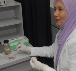
How this is accomplished is by attaching an electrode to the vagus nerve. The other end of the electrode goes to the monitor. The electrode inserted in the throat, touching the vocal cord, is periodically stimulated.  This will alert the surgeon if he is too close for comfort to the RLN and might unintentionally harm it. This gives him ample warning to reassess how he wants to maneuver the dissection.
Dr Rohaizak, said based on statistic 3 out of 100 patients who  undergo thyroid surgery face risk of getting their RLN injured.
He jokingly said after performing such surgery that this actually proves the existence of God. The RLN has a different position than other nerves, this fact has been used by people to argue that humans were  created by God.
“The reason why a surgery is successful before the use of this system is not because the surgeon is great but it is God that protects the patients RLN from human error,” he said.
Risk of Thyroid surgery
Having a husky voice may not be a big deal to many but it can create problems when your voice is the bread winner.
Imagine if you work as teacher, lecturer, or singer; it can mean the end of your career.
A lot of people are reluctant to undergo thyroid surgery because after the surgery they may end up with a different problem.
Imagine coming out the operating theater with a different voice or no voice at all.
In some cases the patient would need a tracheostomy where a hole had to be drilled on the throat to facilitate breathing.
Because of the damage to the RLN the vocal cord loses its ability to automatically shut.
So when you eat the food would fall into your air valve causing you to choke.
These are the risks of thyroid surgery in extreme cases. With the aid of APS in thyroid surgery all these can be avoided.

Creative Surgery
The beauty of this technique is that, it does not require  major investment on the part of the hospital to buy new equipment. The equipment needed is already available in most hospitals. It is used in other nerve surgery like spinal surgery and parotid surgery.
But it only requires minor adjustment to the setting of the machine. Then it will become a revolutionary safety instruments for patients in a thyroid surgery.
It does however need an electrode which is connected to the vagus nerve in the neck.
The RLN is a branch of the vagus nerve and once the electrode is connected, the screen will display the safe range for surgeons to operate.
The surgeon then would perform the gland extraction guided by the monitor and the beeping alarm sound. It helps the surgeon in doing his work, because he does not have to stop and identify the location of the RLN every minute.
He can focus on removing the gland without worrying he might cut the RLN. As the equipment is readily available, there is no extra cost needed.
But Dr Rohaizak believes if the surgery is implemented as a teaching program in UKM he is confident that he can discuss with the suppliers to bring the cost further down.
This new procedure is a great improvement in terms of safety to the patients. It does not require any investment so any major hospital can do it.
He is even willing to set up a training program. He does not know how hospitals in the country will respond to this new method. What is for sure, he had already received requests from surgeons from other parts of Malaysia to learn about the technique. Next is to have training workshop in the neighbouring countries.

Thursday, May 2, 2013

Malaysia can now create human organs


BANGI,  28 July 2010. After successfully culturing human skin tissue using
 tissue engineering technology, UKM researchers are now going inside the 
human body trying to create other organs.

Some of the organs that they have began culturing is the heart, bone, 
cartilage, respiratory epithelium (wind pipe), nerve, conjuctiva and even
 cornea that covers the eye. They hope to make human tissues and organs
 or at the very least find a way to aid the repair of tissues or organs with
 regenerative medicine.
Professor Dr Ruszymah Idrus, Head of Tissue Engineering Centre at UKM Medical Centre, when explaining the work that is being carried out at the Centre said:
“Our dream is to make tissues or organs like kidney, liver and heart. These are organs that cannot regenerate
 and if they failed, the patient will be very sick and die. We started with cartilage cells and then skin cells., We 
began with the cells that can be cultured relatively easier. After that we move on to more difficult cells.”
The research in tissue engineering started in 1999 in UKMMC when UKM researchers  first started to 
engineer human cartilage. Dr Ruszymah’s husband Dr Aminuddin Saim who is an Ear, Nose and Throat (ENT)
 Surgeon came across a number of infants being born without an ear.
So they began to replicate an experiment which was done in Harvard Medical School where they both had 
Post-Doctoral Training. Human chondrocytes was successfully cultured and reconstructed into a 3-D ear lobe with
 the help of a scaffold. The experiment in Harvard did not use human cells. The implant was inserted at the back 
of a nude mice. A nude mice is an animal that do not reject human cells. Amazingly the human ear matured and
 feels like a normal ear lobe.  
“It is a small one because we wanted to test the technique whether it works, That was human ear at the back o
f the mouse made from human cartilage cells” said Dr Ruszymah.
To appreciate what regenerative medicine is all about and how it can lead to replacing body parts, some 
understanding about this relatively new medical segment of tissue engineering is necessary.
Understanding Tissue engineering
The field of tissue engineering began when knowledge about 
cells and how they behave were known. In 1838-1839,
 Schleiden and Schwann formulated the so-called "Cell Theory" 
based on their microscopic findings. The theory dictates that
 cell retains a dual existence as a distinct entity and as a building
 block in the construction of organisms. 
Analogically, cells are like employees working in a corporate
 entity. The company make things happen but it is the individuals who keep the smooth running of the company. Just think of the employees as human cells and the company is 
the human body
 If a company wants a winning position in the industry, it must have good and competent employees. This can happen by sending the workers out for training nurturing them
 to work towards the company’s goal instead of against it. 
This is what tissue engineering is all about, sending cells to
 team building camp so they would work towards better health for the human body. 
What this completely new segment of tissue engineering does to a patient is it gives them shorter recovery period
 and less traumatic medical treatment. Imagine a person who needs a knee replacement, instead of having his
 knee cut open and replaced by foreign object, all he will need is an injection and his kneecap will grow back again.
Manufacturing human organs
Similarly, ever since that discovery of the cell theory, scientists have been intrigue with the notion whether 
human cell can be trained outside the human body, pretty much like employees sent by their companies to a team
 building camp. When they discovered that it is possible to do so; to train or manipulate cell, outside the human 
body, now they are taking it a step further. 
“Tissue engineering is about making human body parts with their own stem cells, if for instance a person’s cornea
 is damaged, by manipulating the stem cells from the patient, a new cornea can be created to replace 
the damaged cornea and prevent the patient from blindness,” said Dr Ruszymah.
Heart in progress
Unlike other body parts that have their own healing mechanism, the heart however, cannot heal itself. Currently
 the treatment of coronary heart problems is a by-pass surgery which involves harvesting a vein from your 
leg, cutting his chest open, stopping the heart and stitching the vein taken from the leg to the heart, making a new
 highway for blood to flow into the heart. 
UKM researchers are now working on ways to simplify that process with tissue engineering technology. In the 
future repairing a heart blocked by cholesterol and fat will not involve the cutting open of the heart as in present day surgery. 
“In UKM we’ve just started to culture heart cells.  Of course, we do not culture human heart but we are trying it 
on sheep. In the future, if people have a severe heart attack, we can inject their own stem cell, which have been
 treated to the damage site. It can then grow into new heart muscles and blood vessel.”
Instead of harvesting a vein from the patient’s leg, the blood vessels will grow by itself on the heart. 
This will make open heart surgery today look somewhat barbaric.
Cornea
The researchers have also successfully used tissue-engineering technology to make cornea the layer at the 
front of the eye. If the cornea is damaged, it will become opaque and this will prevent light to enter the eye and causes blindness.
Dr Ruzymah said her team has already achieved success in making cornea and they are waiting to do clinical
 testing before they can bring it to the bed side.
The difference of the cornea tissue engineered by UKM researchers is that they are fully autologous.
“Though there are many places in the world which make cornea but this is fully autologous. The problem with 
autologous engineered tissue is the problem of tissue delivery, it’s difficult to transfer from one geographical
 area to another. Although there are other centres making cornea, the product is not available to our Malaysian 
population.” said Dr Ruszymah.
Cartilage and Bones
Experiments on animals also prove  to be successful for knee joint. This is especially a blessings for osteoathritis 
patients who suffer from debilatating joint pains.
The pain is caused, by damage cartilage in the knee, basically the part that connects the upper leg and the
 lower leg. So when the knee joint rub against each other it causes immense pain. The present treatment is knee
 replacement.
“We’ve also investigated focal defect and osteoarthritis of the knee joint in sheep. The knee joint has a tendency
 to be damaged in sports injury and as we grow old. These injuries can be treated with stem cells injection. 
We’ve proven this in our animal model and the next step is human clinical trial.” said Dr Ruszymah.
What it means really is that the knee joint can be grown back, instead of having to go for a knee replacement.
“ We can also treat non union (bone that does not heal after a fracture with the normal treatment) bones using
 tissue engineering technology, we can connect the bone,” she said.When a bone is broken the present method is  doctors will assist the bone with steel plate 
allowing the broken bones to unite. But using the tissue engineering technology formulated by UKM
 researchers non-union fractured bones can be reconnected again.

Patient wakes up during brain surgery

By Shahfizal Musa
Pix UKMMC


KUALA LUMPUR, 12 March - The National University of Malaysia (UKM) achieved yet another milestone when a patient was made to wake up during a brain surgery last February.

It was UKM Medical Centre first Awake Craniotomy, a procedure to remove brain tumor with minimal or no brain damage to the patient.

Some might think that waking up during surgery is a terrifying experience but in this case it was intentional. The patient needed to be conscious to monitor and preserve his brain functions. The procedure was performed by UKM Neurosurgeon, Associate Prof Dr Ramesh Kumar assisted by three other surgeons, Dr Toh Charng Jeng,  Dr Sanmugarajah Paramasvaran and Dr Ainul Jaffar.

Dr Esa Kamuruzaman was the anesthetist responsible in sedating the patient before his skull was opened up and  for bringing the him to consciousness during the surgery, just before the tumor is cut out.

The complex surgery is commonly done to remove tumors that grow in the eloquent brain. These are areas of the brain that allow us to communicate, perceive, interact and have movement. They regulate our senses, movement and speech, a very important part of living a normal life.

What is fascinating about the unique procedure is the patient participated in his surgery by giving feedback to the surgeon, who in turn will react accordingly. It is quite a scene when the patient woke up during the operation and starts responding to questions. He also gave a priceless grin when he was asked to smile. 

When asked why a patient need to be awake during the surgery, Dr Ramesh explained: “This technique was developed to allow maximum excision of the lesion while causing minimum or no injury to the surrounding normal brain tissue so that the patient has minimal or no functional loss.

“The best way to know that the patient has his functions intact is from his response. You cannot know this when the patient is unconscious”.

Dr Ramesh said the patient will be asked to perform simple task related to the critical area, while the tumor is removed. Any changes in function are monitored and the surgeon stops further dissections in the related area if any adverse effect is detected.

Though current scanning technology like MRI can pin point where the tumor is but it is not accurate enough.

“By resorting only to the MRI scan, the surgery will be very much like walking in a mine field. Any damage to eloquent areas even by a millimeter can mean living with a disability for the rest of the patient’s life. It might harm the ability to speak or move or even think.
“Although functional MRI can show the motor and speech areas, it may not accurately pin point the most important areas and therefore the brain mapping done with direct electrical stimulation on the brain while the patient is awake remains as the most reliable avenue for neurological preservation,” Dr Ramesh said, the patient is given general anesthesia before commencing surgery to render him unconscious. Then the surgeon will get to work and start drilling his skull to gain access to the patient’s brain.

The sedation is given initialy because the scalp and pericranium (tissue overlying the bone) is pain sensitive while the brain is not.

“Once the skull is opened the anesthesia is reversed and the patient is awakened. Upon completion of the excision of the lesion, the patient is put to sleep again and the skull opening is then closed”.

It is no secret that Awake Craniotomy is a very scary option to the patient so before the patient undergo the procedure he is counselled and guided every step of the way.
The patient is now recovering splendidly. He is fit enough to give a press conference to the media.