 BANGI, 28 July 2010. After successfully culturing human skin tissue using
tissue engineering technology, UKM researchers are now going inside the
human body trying to create other organs.
Some of the organs that they have began culturing is the heart, bone,
cartilage, respiratory epithelium (wind pipe), nerve, conjuctiva and even
cornea that covers the eye. They hope to make human tissues and organs
or at the very least find a way to aid the repair of tissues or organs with
regenerative medicine.
cartilage, respiratory epithelium (wind pipe), nerve, conjuctiva and even
cornea that covers the eye. They hope to make human tissues and organs
or at the very least find a way to aid the repair of tissues or organs with
regenerative medicine.
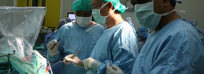
Professor Dr Ruszymah Idrus, Head of Tissue Engineering Centre at UKM Medical Centre, when explaining the work that is being carried out at the Centre said:
“Our dream is to make tissues or organs like kidney, liver and heart. These are organs that cannot regenerate
and if they failed, the patient will be very sick and die. We started with cartilage cells and then skin cells., We
began with the cells that can be cultured relatively easier. After that we move on to more difficult cells.”
and if they failed, the patient will be very sick and die. We started with cartilage cells and then skin cells., We
began with the cells that can be cultured relatively easier. After that we move on to more difficult cells.”
The research in tissue engineering started in 1999 in UKMMC when UKM researchers first started to
engineer human cartilage. Dr Ruszymah’s husband Dr Aminuddin Saim who is an Ear, Nose and Throat (ENT)
Surgeon came across a number of infants being born without an ear.
engineer human cartilage. Dr Ruszymah’s husband Dr Aminuddin Saim who is an Ear, Nose and Throat (ENT)
Surgeon came across a number of infants being born without an ear.
So they began to replicate an experiment which was done in Harvard Medical School where they both had
Post-Doctoral Training. Human chondrocytes was successfully cultured and reconstructed into a 3-D ear lobe with
the help of a scaffold. The experiment in Harvard did not use human cells. The implant was inserted at the back
of a nude mice. A nude mice is an animal that do not reject human cells. Amazingly the human ear matured and
feels like a normal ear lobe.
“It is a small one because we wanted to test the technique whether it works, That was human ear at the back o
f the mouse made from human cartilage cells” said Dr Ruszymah.
f the mouse made from human cartilage cells” said Dr Ruszymah.
To appreciate what regenerative medicine is all about and how it can lead to replacing body parts, some
understanding about this relatively new medical segment of tissue engineering is necessary.
understanding about this relatively new medical segment of tissue engineering is necessary.
Understanding Tissue engineering
The field of tissue engineering began when knowledge about
cells and how they behave were known. In 1838-1839,
Schleiden and Schwann formulated the so-called "Cell Theory"
based on their microscopic findings. The theory dictates that
cell retains a dual existence as a distinct entity and as a building
block in the construction of organisms.
cells and how they behave were known. In 1838-1839,
Schleiden and Schwann formulated the so-called "Cell Theory"
based on their microscopic findings. The theory dictates that
cell retains a dual existence as a distinct entity and as a building
block in the construction of organisms.
Analogically, cells are like employees working in a corporate
entity. The company make things happen but it is the individuals who keep the smooth running of the company. Just think of the employees as human cells and the company is
the human body
entity. The company make things happen but it is the individuals who keep the smooth running of the company. Just think of the employees as human cells and the company is
the human body
If a company wants a winning position in the industry, it must have good and competent employees. This can happen by sending the workers out for training nurturing them
to work towards the company’s goal instead of against it.
This is what tissue engineering is all about, sending cells to
team building camp so they would work towards better health for the human body.
to work towards the company’s goal instead of against it.
This is what tissue engineering is all about, sending cells to
team building camp so they would work towards better health for the human body.
What this completely new segment of tissue engineering does to a patient is it gives them shorter recovery period
and less traumatic medical treatment. Imagine a person who needs a knee replacement, instead of having his
knee cut open and replaced by foreign object, all he will need is an injection and his kneecap will grow back again.
and less traumatic medical treatment. Imagine a person who needs a knee replacement, instead of having his
knee cut open and replaced by foreign object, all he will need is an injection and his kneecap will grow back again.
Manufacturing human organs
Similarly, ever since that discovery of the cell theory, scientists have been intrigue with the notion whether
human cell can be trained outside the human body, pretty much like employees sent by their companies to a team
building camp. When they discovered that it is possible to do so; to train or manipulate cell, outside the human
body, now they are taking it a step further.
human cell can be trained outside the human body, pretty much like employees sent by their companies to a team
building camp. When they discovered that it is possible to do so; to train or manipulate cell, outside the human
body, now they are taking it a step further.
“Tissue engineering is about making human body parts with their own stem cells, if for instance a person’s cornea
is damaged, by manipulating the stem cells from the patient, a new cornea can be created to replace
the damaged cornea and prevent the patient from blindness,” said Dr Ruszymah.
is damaged, by manipulating the stem cells from the patient, a new cornea can be created to replace
the damaged cornea and prevent the patient from blindness,” said Dr Ruszymah.
Heart in progress
Unlike other body parts that have their own healing mechanism, the heart however, cannot heal itself. Currently
the treatment of coronary heart problems is a by-pass surgery which involves harvesting a vein from your
leg, cutting his chest open, stopping the heart and stitching the vein taken from the leg to the heart, making a new
highway for blood to flow into the heart.
the treatment of coronary heart problems is a by-pass surgery which involves harvesting a vein from your
leg, cutting his chest open, stopping the heart and stitching the vein taken from the leg to the heart, making a new
highway for blood to flow into the heart.
UKM researchers are now working on ways to simplify that process with tissue engineering technology. In the
future repairing a heart blocked by cholesterol and fat will not involve the cutting open of the heart as in present day surgery.
future repairing a heart blocked by cholesterol and fat will not involve the cutting open of the heart as in present day surgery.
“In UKM we’ve just started to culture heart cells. Of course, we do not culture human heart but we are trying it
on sheep. In the future, if people have a severe heart attack, we can inject their own stem cell, which have been
treated to the damage site. It can then grow into new heart muscles and blood vessel.”
on sheep. In the future, if people have a severe heart attack, we can inject their own stem cell, which have been
treated to the damage site. It can then grow into new heart muscles and blood vessel.”
Instead of harvesting a vein from the patient’s leg, the blood vessels will grow by itself on the heart.
This will make open heart surgery today look somewhat barbaric.
This will make open heart surgery today look somewhat barbaric.
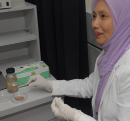
Cornea
The researchers have also successfully used tissue-engineering technology to make cornea the layer at the
front of the eye. If the cornea is damaged, it will become opaque and this will prevent light to enter the eye and causes blindness.
front of the eye. If the cornea is damaged, it will become opaque and this will prevent light to enter the eye and causes blindness.
Dr Ruzymah said her team has already achieved success in making cornea and they are waiting to do clinical
testing before they can bring it to the bed side.
testing before they can bring it to the bed side.
The difference of the cornea tissue engineered by UKM researchers is that they are fully autologous.
“Though there are many places in the world which make cornea but this is fully autologous. The problem with
autologous engineered tissue is the problem of tissue delivery, it’s difficult to transfer from one geographical
area to another. Although there are other centres making cornea, the product is not available to our Malaysian
population.” said Dr Ruszymah.
autologous engineered tissue is the problem of tissue delivery, it’s difficult to transfer from one geographical
area to another. Although there are other centres making cornea, the product is not available to our Malaysian
population.” said Dr Ruszymah.
Cartilage and Bones
Experiments on animals also prove to be successful for knee joint. This is especially a blessings for osteoathritis
patients who suffer from debilatating joint pains.
patients who suffer from debilatating joint pains.
The pain is caused, by damage cartilage in the knee, basically the part that connects the upper leg and the
lower leg. So when the knee joint rub against each other it causes immense pain. The present treatment is knee
replacement.
lower leg. So when the knee joint rub against each other it causes immense pain. The present treatment is knee
replacement.
“We’ve also investigated focal defect and osteoarthritis of the knee joint in sheep. The knee joint has a tendency
to be damaged in sports injury and as we grow old. These injuries can be treated with stem cells injection.
We’ve proven this in our animal model and the next step is human clinical trial.” said Dr Ruszymah.
to be damaged in sports injury and as we grow old. These injuries can be treated with stem cells injection.
We’ve proven this in our animal model and the next step is human clinical trial.” said Dr Ruszymah.
What it means really is that the knee joint can be grown back, instead of having to go for a knee replacement.
“ We can also treat non union (bone that does not heal after a fracture with the normal treatment) bones using
tissue engineering technology, we can connect the bone,” she said.When a bone is broken the present method is doctors will assist the bone with steel plate
allowing the broken bones to unite. But using the tissue engineering technology formulated by UKM researchers non-union fractured bones can be reconnected again.